


Top>Research>Building plastic models (Plamodel) of cells
 Index
Index
Hiroaki Suzuki [profile]
Education Course
Building plastic models (Plamodel) of cells
Hiroaki Suzuki
Associate Professor, Faculty of Science and Engineering, Chuo University
Areas of Specialization: Micro/Nano Biodevices and Biophysics
Models
When I was in elementary school, plastic models (or plamodel in short) of Mobile Suit Gundam were extremely popular. I loved Gundam myself. During my last few years of elementary school, I often drew pictures of Gundam and other mobile suits by heart. Plastic models are models which were made from plastics. Now, what exactly is a model? A model can be defined as an approximate replica of the original. Today, a life-size Gundam can be found in Odaiba. However, it is impossible for people to actually operate and drive that Gundam, so it is nothing more than a life-size model.
Plastic Gundam models are nothing more than a hobby; the models are not used to any benefit. However, there are many “useful models” in our world. For example, when designing an airplane or car, a model is used in wind tunnel tests to evaluate the aerodynamic properties of the design. Computer simulations of physical phenomena are also another type of model. When designing an airplane or car, computer simulations for the flow of fluids around the object provide a highly accurate evaluation even without conducting an actual physical experiment. This type of computation model makes evaluation possible by replicating the actual physical phenomena.
Bio-models and cell models
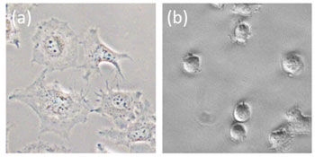
Figure 1: Cells cultivated in our laboratory. (a) Adherent HeLa cells and (b) non-adherent Jurkat cells. The size of the most animal cells ranges from 10 to 50 microns.
Now, to change the subject, I would like to discuss models of organisms (living creatures). A short time ago, the robot dog AIBO achieved great popularity. AIBO can also be called a model. Instead of being a faithfully replication of a dog, AIBO copies (models) how a dog moves and interacts with humans. In my department, there is a professor creating robots which model the movement of worms and water striders. These models are expected to fulfill a variety of roles ranging from medical treatment to space exploration. Delving even further into the subject, I would now like to discuss models of an even smaller scale. Cells are known as the smallest unit of life (Figure 1). DNA, proteins and other molecules inside of cells are not considered as living. However, cells move around and divide, so many people consider them to be alive. Today, great advancements have been made in micro and nano science and technology (otherwise known as nano-technology). Have theses advancements made it possible to create models of cells?
Substance of cells
Cells have an extremely complicated structure. Anyone who has read a biology textbook for junior high school or high school has seen an illustration explaining the contents of cells. All such illustrations show chromosomes inside of the nuclear membrane, surrounded by the endoplasmic reticulum and Golgi body. The cytoplasm is shown to contain mitochondria and lysosomes. Of course, creating such a complicated structure would be difficult and impossible (Note 1). Now, let’s consider this problem from the perspective of models. A model is created by copying certain characteristics of the original. It does not have to be an exact replica of the actual object. For example, when using an aircraft model in wind tunnel tests, it is sufficient to replicate the exterior shape of the aircraft. There is no need to replicate the equipment, control devices or passenger seats within the aircraft. In the case of a cell model, what elements need to be replicated to produce an approximate copy? A cell can be roughly described as DNA and proteins packed into a bag known as the cell membrane.
Method of creating a cell model
Actually, a model of the cell membrane which forms the exterior shape of a cell can be made relatively easily even as a high school level experiment. The cell membrane is made from phospholipids, which are molecules resembling soap molecules. A single lipid molecule is composed of a hydrophilic moiety (water loving) and a hydrophobic moiety (water repelling). Therefore, models use a lipid bilayer in which the hydrophobic moiety face each other without contacting water (Figure 2a). In practice, dried film of phospholipids extracted from egg yolks and soy beans are made in a flask. Simply adding water and waiting produces the wondrous result of numerous objects shaped like balls and long, thin balloons (Figure 2b). This process is about a simple as making instant soup from freeze-dried ingredients. In laboratories, a slightly more sophisticated method is used to create a large number of cell membrane models (known as liposomes) shaped like almost perfect balls (Figure 2c). By simply adding the extracted or synthesized DNA and proteins in the water (the hot water for instant soup), these molecules enter the emerging liposomes. The result is a simple cell model.
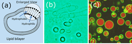
Figure 2a: Schematic diagram of a cell membrane model (liposomes).
Figure 2b: Liposomes approximately the same size as a cell. Made during an experiment by new students in our laboratory.
Figure 2c: Spherical liposomes made in our laboratory. Dyed with fluorescent markers for easy visibility.
Model of cell function
I have explained how to create rough copies of cells. Just like an aircraft exterior in wind tunnel tests, liposomes can be used to investigate the properties of cell membrane, to examine changes in shape, and to observe interaction between the membrane and other substances. However, scientists are not satisfied with such a rough copy. Liposomes are not even close to a real living cell in that they are not alive (no metabolism) and do not move.
In the 1990s, a research group including members from Japan succeeded in synthesizing proteins inside liposomes by encapsulating DNA, proteins and other solutions inside the liposomes[1]. In real cells, proteins are synthesized based on the information written in DNA to be alive. Thus, this was a major scientific advance in creating a cell model. However, this cell model does not acquire food from the outside. Therefore, protein synthesis stops after a few hours. To solve this problem, a different research group created a protein which opens a tiny hole in the model membrane. This enabled nutrition from outside the membrane to enter the liposomes and protein synthesis continues for a longer period of several days[2].
Model of proliferating cells
Although liposomes which continue to synthesize protein seem to be alive, they leave something to be desired because they do not move even the slightest bit. Actual cells incorporate nutrition and proliferate rapidly. In other words, they produce almost exact copies of themselves. Can such proliferation be achieved through cell models?
Theoretically, this is now somewhat possible. A research group led by Dr. Jack Szostak, winner of the 2009 Nobel Prize in Physiology or Medicine, demonstrated that adding fatty acid molecules to a cell membrane model made from fatty acids causes the model to rapidly stretch and that the model breaks into approximately the same size when perturbation is added[3]. A different research group led by Professor Sugawara from the University of Tokyo demonstrated that a cell membrane model made from chemically-synthesized amphipathic molecules divides when the model enlarges due to acquiring membrane components from the outside[4]. A research group to which I belong has demonstrated how a cell model divides due to an extremely simple physical effect. Our findings were published in 2012. Cells have cell membranes which are packed with DNA, proteins and other components. In our research, replicating that condition (packing macromolecules at high density) caused a change in which the model divides (Figure 3)[5].
(Video: http://youtube/sY64KXAexIA![]() , http://youtube/OCqvxpDAo-o
, http://youtube/OCqvxpDAo-o![]() )
)
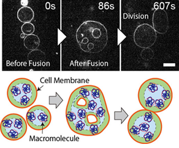
Figure 3: An artificially created cell model divides spontaneously. After the fusion of multiple liposomes, a change occurs which leads to spontaneous division even without external stimulation.
Cell models & beyond
I belong to the Department of Precision Mechanics. Some people may wonder why we are conducting such cellular research in the field of mechanics. Returning to my introductory example of plastic models, the same method of thinking applies when creating something hard like a machine or something soft like a cell. Furthermore, there have been significant advancements in micro-processing and micro-machines. It is now relatively easy to create mechanical structures of roughly the same size as cells and proteins. Although not discussed in this article, my laboratory is creating microscopically precise machines (structures) for analyzing individual cells, as well as for reconstructing cell membrane on microchips. We are approaching an age in which micro and nano technology can be used to directly operate cells, DNA and proteins. As such, there are high expectations for further contributions by mechanical engineers.
What are the benefits of artificially created cell models? Unfortunately, there are almost no examples of uses which provide immediate benefits. Research for creating cell models and artificial cells is conducted as fundamental research and as a challenge to mankind. However, ten or twenty years in the future, there may come a day in which cell models can be used to produce pharmaceuticals or to contribute to the solution of food and energy issues. Someday, cell models might also be used as educational material for science classes in elementary school or junior high school. I hope that everyone will remember that there is a unique form of research that involves creating cell models. Personally, I hope to use a 3D printer to create a plastic model of a cell.
(Note 1) Of course, bacteria and viruses are much simpler structures. However, I am limiting the scope of discussion to make this article easily understood to novices.
[1] Nomura et al., Biophysics, 48, 174, 2008. [2] Noireaux & Libchaber, PNAS, 101, 17669, 2004. [3] Zhu & Szostak, JACS, 131, 5705, 2009. [4] Kurihara et al., Nature Chemistry, 3, 775, 2011. [5] Terasawa et al., PNAS, 109, 5942, 2012.
- Hiroaki Suzuki
Associate Professor, Faculty of Science and Engineering, Chuo University
Areas of Specialization: Micro/Nano Biodevices and Biophysics - Hiroaki Suzuki was born in Aichi Prefecture in 1973. He graduated from the Faculty of Engineering, the University of Tokyo in 1997.
In 1999, he completed the Master’s Program in the Department of Mechanical Engineering at the Graduate School of Engineering, the University of Tokyo.
In 2003, he completed the Doctoral Program in the Department of Mechanical Engineering at the Graduate School of Engineering, University of Tokyo. He holds a PhD in engineering.
He served as Assistant Professor at the Institute of Industrial Science, the University of Tokyo and Associate Professor at the Graduate school of Information Science and Technology, Osaka University before assuming his current position in 2013.
His current research themes include bio-chips, cell models and engineering for self-organization.

- Research Activities as a Member of Research Fellowship for Young Scientists (DC1), Japan Society for the Promotion of Science (JSPS) Shuma Tsurumi
- Important Factors for Innovation in Payment Services Nobuhiko Sugiura
- Beyond the Concepts of Fellow Citizens and Foreigners— To Achieve SDGs Goal 10 “Reduce Inequality Within and Among Countries” Rika Lee
- Diary of Struggles in Cambodia Fumie Fukuoka
- How Can We Measure Learning Ability?
—Analysis of a Competency Self-Assessment Questionnaire— Yu Saito / Yoko Neha - The Making of the Movie Kirakira Megane